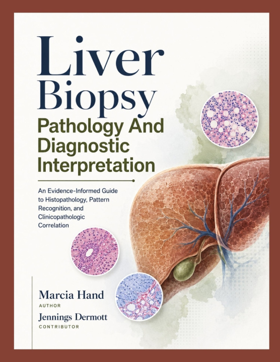
A small tissue core can carry an enormous diagnostic burden. This reference helps you turn limited liver specimens into clear, defensible, clinically meaningful conclusions.
Liver Biopsy Pathology and Diagnostic Interpretation presents a practical, evidence-informed approach to hepatic tissue assessment-from specimen adequacy and normal microanatomy to inflammatory, cholestatic, steatotic, vascular, metabolic, transplant, and neoplastic disease. Rather than reducing interpretation to isolated microscopic features, the book shows you how to identify the dominant injury pattern, build a prioritized differential, select high-value ancillary studies, and communicate uncertainty without losing clinical usefulness.
Designed for practicing pathologists, pathology residents and fellows, hepatology professionals, laboratory teams, and advanced clinical trainees, this comprehensive text follows a specimen-to-pattern-to-etiology-to-report sequence. It connects morphology with laboratory trends, imaging, medication timelines, immune status, transplant history, previous tissue, and molecular findings. The result is a disciplined framework that remains useful when the biopsy is fragmented, treated, heterogeneous, near a diagnostic threshold, or discordant with the clinical impression.
Inside, you will learn to:
- Evaluate core length, portal tract representation, fragmentation, orientation, and sampling limitations.
- Distinguish portal, lobular, interface, steatotic, cholestatic, vascular, granulomatous, fibrosing, and infiltrative patterns.
- Select and interpret trichrome, reticulin, iron, PAS-D, copper-related stains, immunohistochemistry, in situ hybridization, and molecular tests.
- Separate active injury from chronic fibrosis, collapse, treatment effect, regression, and advanced burned-out disease.
- Approach viral, autoimmune, drug-related, biliary, metabolic, pediatric, allograft, benign lesion, primary tumor, metastatic, and hematolymphoid specimens.
- Translate microscopic findings into structured reports that state evidence, confidence, limitations, and the most relevant next-step correlation.
Integrated cases, reporting checkpoints, diagnostic traps, chapter summaries, and visual teaching figures reinforce the reasoning process at the point where interpretive errors most often occur. Dedicated discussions of tissue stewardship, digital pathology, ancillary-test selection, quality assurance, second opinions, and multidisciplinary review extend the book beyond recognition toward real-world sign-out practice.
Build a more systematic approach to hepatic tissue review and strengthen the clarity, consistency, and clinical value of every report.